Selected papers (see entire list here)
A change in cis-regulatory logic underlying obligate versus facultative muscle multinucleation in chordates
Christopher J. Johnson, Zheng Zhang, Haifeng Zhang, Renjie Shang, Katarzyna M. Piekarz, Pengpeng Bi, and Alberto Stolfi (2024)
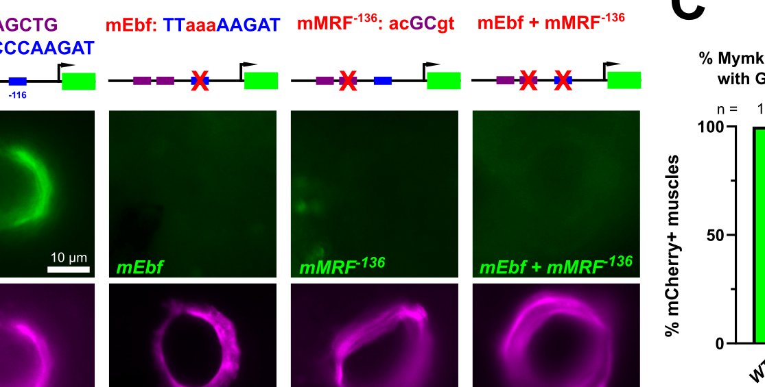
Tunicates and vertebrates both have multinucleated muscles that form through fusion directed by the fusogenic transmembrane protein Myomaker, which evolved through gene duplication and neofunctionalization in their last common ancestor. Where they diverge is in the mode of muscle fusion and multinucleation- while vertebrates activate the expression of Myomaker in all muscles, in tunicates its expression is limited to post-metamorphic muscles. As a result, all skeletal muscles in vertebrates are multinucleated, while in tunicates muscle multinucleation is facultative, i.e. selectively deployed. Here we show the genetic basis for this difference. In vertebrates, MRF factors alone are sufficient to activate the expression of Myomaker. In tunicates, a combination of MRF and Ebf factors is necessary and sufficient. We show that this combinatorial logic is likely encoded in cis, and that manipulating MRF binding site affinities is sufficient to switch this logic to a more vertebrate-like one that requires only MRF. LINK TO PREPRINT
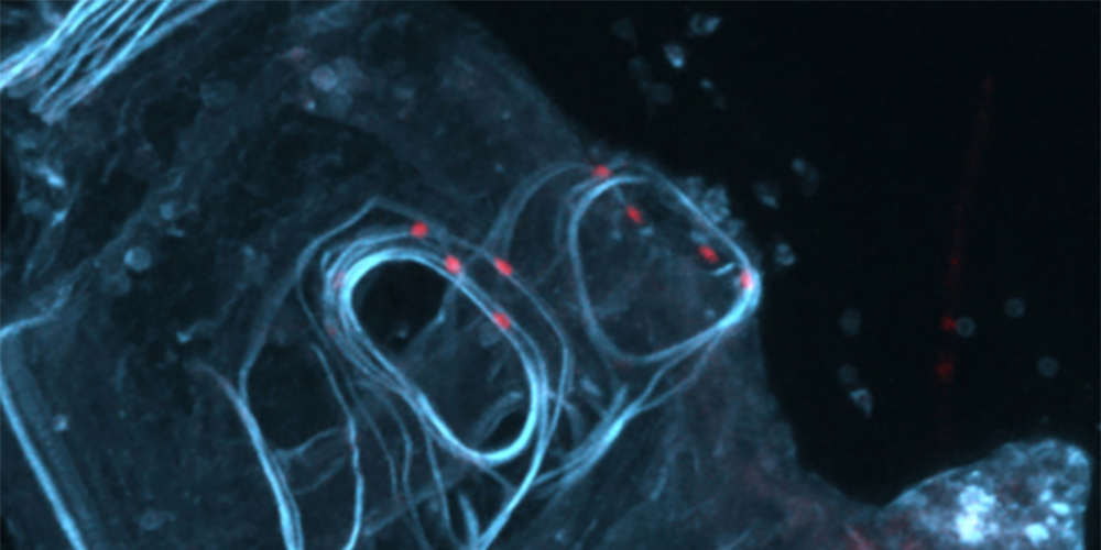
Specification of distinct cell types in a sensory-adhesive organ important for metamorphosis in tunicate larvae
Christopher J. Johnson*, Florian Razy-Krajka*, Fan Zeng, Katarzyna M. Piekarz, Shweta Biliya, Ute Rothbächer, and Alberto Stolfi (2024)
*equal contributions
The sensory/adhesive papillae are a set of organs comprised of highly organized arrays of different cells types that are crucial for the attachment and metamorphosis of the Ciona larva. Using tissue-specific CRISPR/Cas9-mediated mutagenesis and other molecular perturbations, we revealed the roles of key transcription factors and signaling pathways that are important for patterning and shaping these papillae, producing the adhesive glue that allows for larval attachment during settlement, and for triggering the onset of metamorphosis. LINK TO UNCORRECTED PROOF
A gene regulatory network for specification and morphogenesis of a Mauthner Cell homolog in non-vertebrate chordates
Kwantae Kim, Katarzyna M. Piekarz, and Alberto Stolfi (2024)
The descending decussating neurons (ddNs) of the Ciona larva have been proposed to be the homologs of the the Mauthner Cells of fish and amphibians, on account of their hindbrain origin and position in the larval swimming circuit. Here we dissect in greater detail a gene regulatory network downstream of the key regulator, Pax3/7, operating in the ddNs to drive the expression of effector genes that might underlie the development of their unique morphology. LINK TO PREPRINT
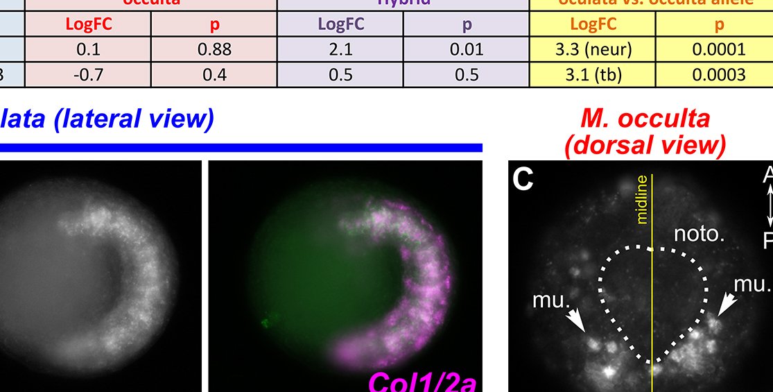
Loss of collagen gene expression in the notochord of the tailless tunicate Molgula occulta
Sydney Popsuj, Anna Di Gregorio, Billie J. Swalla, and Alberto Stolfi (2023)
We have published another paper showing that the evolution of tailless, non-swimming larvae in Molgula occulta is accompanied by the evolutinary loss of tissue-specific expression of key developmental genes. The genes themselves are not lost from the genome (probably because they are important for the adult phase), but rather they have lost key cis-regulatory elements driving their expression in larval cell types that are crucial for swimming larvae, but dispensable for non-swimming ones. In this case, Collagen1/2a, which encodes an extracellular matrix protein that we show is important for proper notochord morphogenesis in the tailed larvae of Ciona. PDF
Specification and survival of post-metamorphic branchiomeric neurons in the hindbrain of a non-vertebrate chordate
Eduardo D. Gigante, Katarzyna M. Piekarz, Alexandra Gurgis, Leslie Cohen, Florian Razy-Krajka, Sydney Popsuj, Hussan S. Ali, Shruthi Mohana Sundaram, and Alberto Stolfi (2023)
Here we investigated the “Neck”, a group of cells situated in between the brain and motor ganglion in the Ciona larva, and that gives rise part of the adult nervous system following metamorphosis. We show using CRISPR/Cas9 and RNAseq approaches that these cells are specified by Pax2/5/8 and that Ephrin/FGF signaling controls the balance of larval vs. post-metamorphic neurons in this region. Suppressing FGF results in loss of adult ciliomotor neurons, likely through converting them into larval neurons that are eliminated during metamorphosis. LINK TO PREPRINT
Using CRISPR/Cas9 to identify genes required for mechanosensory neuron development and function
Christopher J. Johnson, Akhil Kulkarni, William J. Buxton, Tsz Y. Hui, Anusha Kayastha, Alwin A. Khoja, Joviane Leandre, Vanshika V. Mehta, Logan Ostrowski, Erica G. Pareizs, Rebecca L. Scotto, Vanesa Vargas, Raveena M. Vellingiri, Giulia Verzino, Rhea Vohra, Saurabh C. Wakade, Veronica M. Winkeljohn, Victoria M. Winkeljohn, Travis M. Rotterman, and Alberto Stolfi (2023)
We took advantage of the low-cost and simplicity of Ciona by using tissue-specific CRISPR/Cas9-mediated mutagenesis to screen for genes potentially involved in mechanosensation and metamorphosis, in the context of an undergraduate “capstone” research course. This small screen revealed at least one gene, Vamp1/2/3, that appears crucial for the ability of the papillae to trigger metamorphosis. We also provided step-by-step protocols and tutorials associated with this course, in the hope that it might be replicated in similar CRISPR-based laboratory courses wherever Ciona are available. All 17 students and the TA are co-authors on the study. PDF
Specification of distinct cell types in a sensory-adhesive organ for metamorphosis in the Ciona larva
Christopher J. Johnson*, Florian Razy-Krajka*, Fan Zeng, Katarzyna M. Piekarz, Shweta Biliya, Ute Rothbächer, and Alberto Stolfi (2023)
*equal contributions
The sensory/adhesive papillae are a set of organs comprised of highly organized arrays of different cells types that are crucial for the attachment and metamorphosis of the Ciona larva. Using tissue-specific CRISPR/Cas9-mediated mutagenesis and other molecular perturbations, we revealed the roles of key transcription factors and signaling pathways that are important for patterning and shaping these papillae, producing the adhesive glue that allows for larval attachment during settlement, and for triggering the onset of metamorphosis. LINK TO PREPRINT
Pax3/7 regulates neural tube closure and patterning in a non-vertebrate chordate
Kwantae Kim, Jameson Orvis, and Alberto Stolfi (2022)
In this study, we used CRISPR/Cas9-mediated mutagenesis to investigate the roles of Pax3/7 in neural tube closure and patterning in Ciona. We found that Pax3/7 is expressed at different times in distinct lineages that give rise to different cells of the neural tube. At the neurula stage, Pax3/7 in the neural plate borders is essential for neural tube closure, but does not appear to be required for melanin-contianing pigment cell specification. In contrast, Pax3/7 is required for the specification of the Descending Decussating Neuron (ddN) pair of the larval Motor Ganglion. These findings refine our model for the evolution of chordate neurodevelopment. PDF
Evolution of a chordate-specific mechanism for myoblast fusion
Haifeng Zhang, Renjie Shang, Kwantae Kim, Wei Zheng, Christopher J. Johnson, Lei Sun, Xiang Niu, Liang Liu, Jingqi Zhou, Lingshu Liu, Zheng Zhang, Theodore A. Uyeno, Jimin Pei, Skye D. Fissette, Stephen A. Green, Sukhada P. Samudra, Junfei Wen, Jianli Zhang, Jonathan T. Eggenschwiler, Douglas B. Menke, Marianne E. Bronner, Nick V. Grishin, Weiming Li, Kaixiong Ye, Yang Zhang, Alberto Stolfi, and Pengpeng Bi (2022)
Multinucleated vertebrate muscles are formed through a process of cell-cell fusion driven by the transmembrane protein Myomaker and its co-factor Myomixer. Previously thought to be a vertebrate innovation, we found that Myomaker is also expressed in developing multinucleated juvenile muscles in Ciona (but not mononucleated larval muscles), and that knocking it out by CRISPR/Cas9 disrupts juvenile muscle development. This collaborative work ties together work in tunicates and both jawed and jawless vertebrates to reveal the complex evolutionary history of this chordate-specific mechanism for muscle cell fusion. PDF
Ebf activates expression of a cholinergic locus in a multipolar Motor Ganglion interneuron subtype in Ciona
Sydney Popsuj and Alberto Stolfi (2021)
Here we show yet another important role for the transcription factor Ebf (COE, or Collier/Olf/Ebf) in neuronal specification in Ciona. More precisely, we found that the sole cholinergic neuron of the Ascending Motor Ganglion complex (AMG Neuron 5) is specified by Ebf. While all other AMG neurons are GABAergic, AMG5 is cholinergic. This is because it is the only AMG neuron to express Ebf, which directly activates the expression of the conserved cholinergic locus VAChT/ChAT. PDF
A cis-regulatory change underlying the motor neuron-specific loss of Ebf expression in immotile tunicate larvae
Elijah K. Lowe, Claudia Racioppi, Nadine Peyriéras, Filomena Ristoratore, Lionel Christiaen, Billie J. Swalla, and Alberto Stolfi (2021)
We identified a cis-regulatory change underlying the reduced expression of a key neuronal regulator, Ebf (Collier/Olf/Ebf, or COE) in the Motor Ganglion of tail-less, non-swimming larvae of Molgula occulta. The MG-specific reduction of Ebf expression in turn might underlie a loss of motor neuron differentiation, as a result of relaxed purifying selection. PDF
Expression of smooth muscle-like effectors and core cardiomyocyte regulators in the contractile papillae of Ciona
Christopher J. Johnson, Florian Razy-Krajka, and Alberto Stolfi (2020)
Previous single-cell sequencing from the lab revealed the transcriptional profile of the enigmatic Axial Columnar Cells (ACCs) of the anterior sensory organs known as papillae. Here we revealed that the ACCs of Ciona resemble vertebrate myoepithelial cells and might carry out important contractile and sensory functions during larval attachment and metamorphosis. Furthermore, their expression of transcription factors associated with smooth muscle and cardiomyocytes in vertebrates suggests a close evolutionary link between these different contractile cell types. PDF
Regulation of neurogenesis by FGF signaling and Neurogenin in the invertebrate chordate Ciona
Kwantae Kim*, Susanne Gibboney*, Florian Razy-Krajka, Elijah K. Lowe, Wei Wang, and Alberto Stolfi (2020)
*equal contributions
We showed the roles of FGF signaling and the transcription factor Neurogenin in the specification and differentiation of Bipolar Tail Neurons in Ciona. RNAseq was also used to identify potential effectors of Bipolar Tail Neuron cell behaviors like delamination, migration, and polarization downstream of Neurogenin activity. PDF
Effector gene expression underlying neuron subtype-specific traits in the Motor Ganglion of Ciona
Susanne Gibboney*, Jameson Orvis*, Kwantae Kim, Christopher J. Johnson, Paula Martinez-Feduchi, Elijah K. Lowe, Sarthak Sharma, and Alberto Stolfi (2020)
*equal contributions
Transcriptional profiles of two distinct Motor Ganglion neuron subtype (ddN vs. MGIN2) were compared, resulting in candidate effectors of MG neuron subtype-specific functional and morphological traits. Among these are intriguing intracellular and extracellular proteins that might be controlling the dynamic repolarization of ddNs as they extend their axons across the midline. Two candidate effectors (Efcab6-related and Netrin1) were shown, by lineage-specific CRISPR/Cas9-mediated gene knockouts, to be essential for polarized axon outgrowth. PDF
Regulation and evolution of muscle development in tunicates
Florian Razy-Krajka and Alberto Stolfi (2019)
In this review article, we summarize the current literature on muscle specification and differentiation in tunicates, covering all major tunicate classes and orders, including thaliaceans (salps, pyrosomes, and doliolids) and appendicularians. We discuss these findings in the context of tunicate and animal evolution in general. PDF
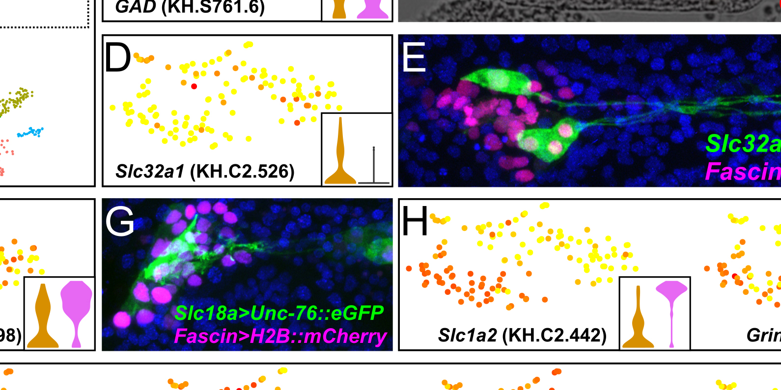
Single-cell transcriptome profiling of the Ciona larval brain
Sarthak Sharma, Wei Wang, and Alberto Stolfi (2019)
Single-cell RNAseq was used to identify specific subsets of cells isolated and selected from the brains of Ciona larvae, and to determine the unique gene expression profiles of each cell type. PDF
Developmental system drift in motor ganglion patterning between distantly related tunicates
Elijah K. Lowe and Alberto Stolfi (2018)
Here we demonstrate that the specification and final arrangement of the distinct neuronal subtypes of the tunicate spinal cord homolog, the Motor Ganglion, is deeply conserved between distantly related solitary tunicates, suggesting that this structure represents an ancient and highly adapted minimal central pattern generator for larval swimming. PDF
Evolutionary loss of melanogenesis in the tunicate Molgula occulta
Claudia Racioppi, Maria Carmen Valoroso, Ugo Coppola, Elijah K. Lowe, C. Titus Brown, Billie J. Swalla, Lionel Christiaen, Alberto Stolfi, and Filomena Ristoratore (2017)
We found that the tunicate Molgula occulta has lost the genes encoding crucial enzymes for melanin synthesis, perhaps because its tail-less larvae do not swim and have no need for pigmented light- and gravity-sensing organs. PDF
Evaluation and rational design of guide RNAs for efficient CRISPR/Cas9-mediated mutagenesis in Ciona
Shashank Gandhi, Maximilian Haeussler, Florian Razy-Krajka, Lionel Christiaen, and Alberto Stolfi (2017)
We developed a novel pipeline for rapid yet accurate design, synthesis, and evaluation of efficient sgRNAs for high-throughput, tissue-specific CRISPR/Cas9 knockouts in Ciona embryos. PDF
Migratory neuronal progenitors arise from the neural plate borders in tunicates
Alberto Stolfi, Kerrianne Ryan, Ian A. Meinertzhagen, and Lionel Christiaen (2015)
We show that a neuronal cell type in the larva of the tunicate Ciona, the bipolar tail neuron, shares a set of features with neural-crest-derived spinal ganglia neurons in vertebrates, hinting at the pre-vertebrate origins of the neural crest. PDF
Tissue-specific genome editing in Ciona embryos by CRISPR/Cas9
Alberto Stolfi, Shashank Gandhi, Farhana Salek, and Lionel Christiaen (2014)
We report the first use of CRISPR/Cas9 for tissue-specific genome editing in the tunicate Ciona. Using this technique to disrupt the Ebf gene in different cell lineages, we revealed that its function is required for specification motor ganglion neurons and atrial siphon muscles. PDF
Divergent mechanisms regulate conserved cardiopharyngeal development and gene expression in distantly related ascidians
Alberto Stolfi*, Elijah Lowe*, Claudia Racioppi, Filomena Ristoratore, C. Titus Brown, Billie J. Swalla, and Lionel Christiaen (2014)
*equal contributions
Comparisons between the distantly related tunicates Molgula and Ciona reveal cryptic turnover
of regulatory mechanisms that underlie identical gene expression patterns. PDF
Coordinated regulation of cholinergic motor neuron traits through a conserved terminal selector gene
Paschalis Kratsios, Alberto Stolfi, Michael Levine, and Oliver Hobert (2012)
We show that UNC-3/Ebf is a terminal selector for cholinergic motor neuron differentiation whose function is conserved across phylogeny, from C. elegans to Ciona. PDF
Genetic and genomic toolbox of the chordate Ciona intestinalis
Alberto Stolfi and Lionel Christiaen (2012)
A review on the tools, techniques, and resources available to the Ciona geneticist, citing examples of studies that employed such strategies in the elucidation of gene function in Ciona: electroporation, transgenesis, genetic screens, and a preview of the emergence of genetic engineering platforms like CRISPR/Cas9. PDF
Neural tube patterning by Ephrin, FGF, and Notch signaling relays
Alberto Stolfi, Eileen Wagner, J. Matthew Taliaferro, Seemay Chou, and Michael Levine (2011)
We show that the Ciona Motor Ganglion is patterned by sequential Ephrin/FGF/MAPK and Delta/Notch signaling events that lead to localized expression of conserved homeodomain codes for the specification of distinct neuronal subtypes. PDF
Neuronal subtype specification in the spinal cord of a protovertebrate
Alberto Stolfi and Michael Levine (2011)
We used fluorescent reporter constructs to label and identify invariant but morphologically distinct moto- and interneuron subtypes in the Motor Ganglion of the Ciona larva. We also perturbed neuronal subtype specification in predictable ways by targeting specific transcription factors and signaling molecules. PDF
Early chordate origins of the vertebrate second heart field
Alberto Stolfi, T. Blair Gainous, John J. Young, Alessandro Mori, Michael Levine, and Lionel Christiaen (2010)
We found that heart progenitor cells of the simple chordate Ciona also generate precursors of the pharyngeal muscles. These precursors express Islet and Tbx1/10, evocative of the splanchnic mesoderm that produces the lower jaw muscles and Second Heart Field of vertebrates. We propose that the last common ancestor of tunicates and vertebrates possessed multipotent cardiopharyngeal muscle precursors, and that their reallocation might have contributed to the emergence of the SHF. PDF
Gene regulatory networks underlying the compartmentalization of the Ciona central nervous system
Kaoru S. Imai, Alberto Stolfi, Michael Levine, and Yutaka Satou (2009)
Here we document provisional gene regulatory networks operating in the developing central nervous system of Ciona, at single-cell resolution. This revealed a striking conservation in neural gene expression and patterning relative to vertebrates. More careful analysis revealed a role for FGF8 in patterning the Ciona hindbrain in a manner reminiscent of the midbrain/hindbrain boundary organizer in vertebrates. PDF